The cleanest answer to peptide vs. protein is “peptides are shorter.” That’s mostly right. The fuller answer adds two more dimensions. How the molecule folds. And what it does in biological systems.
The standard textbook cut-off is roughly 50 amino acids. Below that, biochemists usually call the molecule a peptide. Above it, a protein. The cut-off is a convention, not a hard rule. A more rigorous distinction: proteins reliably fold into stable 3D shapes, peptides usually don’t. Some molecules sit right on the boundary. Insulin (51 residues) gets called a protein. Glucagon (29) and tirzepatide (39) get called peptides. The labeling is shorthand, not biology.
If you’ve seen the same molecule called both a peptide and a small protein in different sources, you weren’t imagining it. The vocabulary really is inconsistent. Insulin gets called both, depending on which textbook you’re reading. The practical distinctions break into three: how long it is, whether it folds, and what it does.
Distinction one: length
The simplest definition uses residue count. The conventions vary slightly by field, but the rough boundaries that show up in most biochemistry textbooks look like this:
- Oligopeptide: 2–10 amino acids. KPV (3 residues), GHK-Cu (3), ipamorelin (5), oxytocin (9), vasopressin (9) all sit here.
- Polypeptide / peptide: 10–50 amino acids. BPC-157 (15), MOTS-c (16), glucagon (29), CJC-1295 (29), tirzepatide (39), retatrutide (39), tesamorelin (44) all fall into this band.
- Small protein: 50–100 amino acids. Insulin (51), GLP-1 precursor fragments, calcitonin (32 but often grouped here), parathyroid hormone (84) sit at this edge.
- Protein: 100+ amino acids. Antibodies, albumin, haemoglobin, most enzymes.
The 50 amino acid cut-off is the most widely cited, but you’ll see 40 in some textbooks and 100 in others. The variability is part of why “peptide” isn’t a regulated term.
Distinction two: folding
A more biologically meaningful distinction: does the molecule fold into a stable 3D shape on its own? Proteins fold. The linear amino acid sequence curls and twists into local helices and sheets, then those elements pack into a final globular or fibrous shape. Larger proteins can also link multiple chains together. The folded shape is what creates a functional binding pocket or active site.
Peptides usually don’t fold reliably on their own. They’re too short. A 10-residue peptide doesn’t have enough sequence to drive stable folding. It floats through many shapes in solution and locks into the one that fits its target only at the moment of binding. Some peptides have local helical or hairpin tendencies (especially in the 20 to 30-residue range), but full 3D folding is generally a protein behavior.
This is why “peptide” can’t be cleanly defined by length alone. Two molecules of the same length can behave very differently. A 30-residue peptide locked by a disulfide bridge folds into a compact, protein-like structure (calcitonin, defensins). A 30-residue linear peptide without that brace floats around like a typical short chain.
The distinction between peptides and proteins is operational rather than fundamental. Both are polymers of L-amino acids, and the boundary used in practice depends on whether the molecule’s function emerges from a stable folded structure or from a flexible linear sequence binding a receptor.
— Lehninger Principles of Biochemistry, 8th edition
Distinction three: function
Proteins typically do work. Enzymes catalyse reactions. Antibodies bind antigens. Structural proteins build cellular scaffolding. Transport proteins shuttle small molecules. The folded shape is the functional unit.
Peptides typically carry signals. They’re messengers between cells. Hormones, neurotransmitters, growth factors, immune signals. They bind targets, trigger reactions, and then get cleared. The flexibility is often a feature, not a bug. A peptide that can flex into multiple shapes can bind multiple targets and tune multiple pathways.
This functional split is where the most useful intuition lives. If a molecule has an active site and turns substrates into products, it’s probably a protein. If it travels from one cell to another and tells the second cell what to do, it’s probably a peptide. There are exceptions to both, but the rule of thumb works for ~80% of cases.
A worked example: tirzepatide
Tirzepatide is a useful test case. It sits right on the fuzzy edge. It’s the active ingredient in Mounjaro and Zepbound. 39 residues, linear, with a fatty chain attached. By length, it’s a peptide. It’s built by chemical synthesis (one residue at a time on a bead, called SPPS), not grown in cells. That’s another peptide hallmark. It binds two cell-surface targets (GIP and GLP-1) as a flexible chain, not as a folded protein domain. Every functional marker says “peptide.”
But its molecular weight is roughly 4,813 g/mol, comparable to small proteins like insulin (5,808 g/mol). And it gets called a “peptide hormone” or “peptide drug” interchangeably. Classification here is settled by convention, not by measurement. Tirzepatide is a peptide because the field has agreed to call it one.
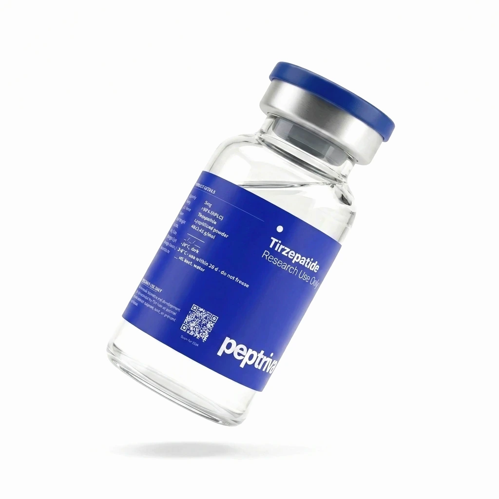
Tirzepatide
A useful boundary case — the largest peptide in our catalog at 39 residues with a C20 fatty acid tail. Dual GIP/GLP-1 receptor agonist with the longest Phase III dataset of any incretin-class compound to date.
How they’re made: another clean split
Manufacturing route is one of the cleanest dividing lines in practice. Peptides up to roughly 50 residues are made chemically using solid-phase synthesis (SPPS). You build the chain one amino acid at a time on a plastic bead. Proteins and very long peptides are produced by growing them in cells. You engineer a DNA sequence into bacteria, yeast, or mammalian cells and let the organism build the protein for you.
This matters because of cost and complexity. SPPS scales decently up to about 50 residues. Beyond that, errors compound. At 99% efficiency per step, a 60-residue chain still ends up only about 55% correct sequence. The rest is deletion or insertion errors. Cell-grown proteins don’t have that scaling problem. Cells produce full-length proteins reliably. But cell production requires genetic engineering, fermentation tanks, downstream purification, and folding validation. Both routes are expensive. They just have different cost structures.
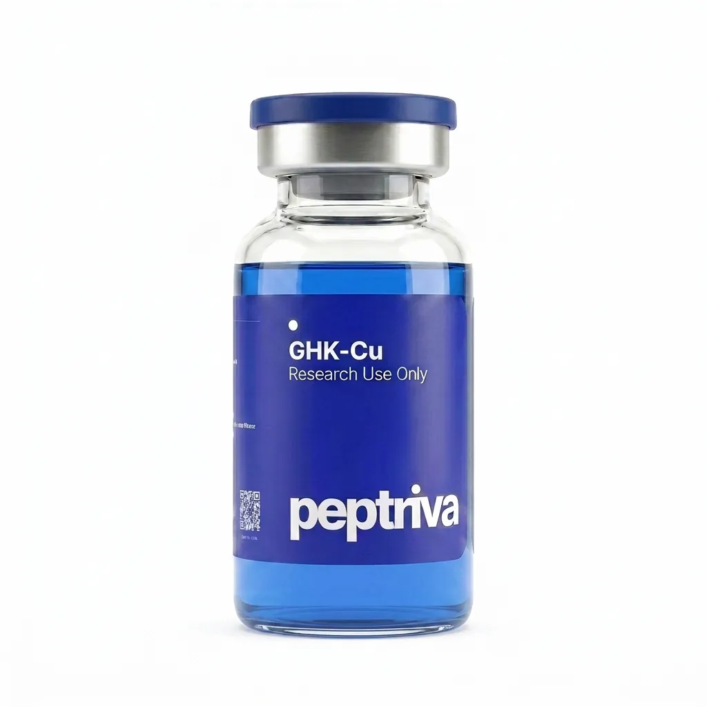
GHK-Cu
A 3-residue copper-binding tripeptide (Gly-His-Lys) — the small-molecule end of the size scale, far below the ~50-residue peptide/protein boundary. Supplied for in-vitro research. COA available with each lot.
Why the distinction matters at all
It matters in three places.
Regulatory. Drug-approval paths for small molecules, peptides, and biologics all differ. Peptides under about 40 residues usually go through the FDA’s small-molecule division. Proteins and antibodies go through the biologics division with different testing rules.
Manufacturing. Chemical synthesis on a bead vs. growing the molecule in engineered cells is essentially a peptide-vs-protein choice. The cost gap is real.
Drug behavior. Peptides break down faster in vivo than proteins do. Proteins’ folded structure shields them from proteolytic enzymes. Most peptide drug design is therefore a clearance-time engineering problem. Most protein drug design is more about manufacturability and immune reactions.
The takeaway. “Peptide” and “protein” are useful shorthand, not biology. The boundary is somewhere around 50 amino acids, but length, folding, function, and manufacturing route all play a role. And the molecules that sit right on the boundary (insulin, GLP-1 drugs, calcitonin) include some of the most useful drugs we have.
Questions worth asking
- How long is it? The most reliable single criterion. Under 50 residues is usually called a peptide. Over 50 is usually called a protein.
- Does it fold? A molecule with a defined 3D structure that’s required for its function is behaving like a protein.
- What does it do? Signal-carrying messengers behave like peptides. Catalytic and structural workhorses behave like proteins.
- How is it made? Chemically synthesized molecules are usually peptides. Cell-grown molecules are usually proteins.
- How is it regulated? Different FDA divisions handle peptides and proteins with different testing rules. The label often decides the approval path.
What to know now
- Common cut-off: ~50 amino acids. Below = peptide; above = protein. The boundary is a convention, not biology.
- Folding: proteins reliably fold into 3D structures; peptides usually don’t fold on their own and adopt their conformation upon receptor binding.
- Function: peptides mostly carry signals between cells; proteins mostly do catalytic or structural work.
- Manufacturing: peptides via SPPS (solid-phase synthesis); proteins via recombinant expression in bacteria, yeast, or mammalian cell lines.
- Edge cases: insulin (51 aa) is called a protein; tirzepatide (39 aa) is called a peptide. The line is fuzzy on purpose.
What we’re watching
One trend worth tracking: the rise of peptide-protein hybrids. Antibody-drug conjugates and peptide-Fc fusions effectively glue a short peptide signalling unit to a long protein backbone (often an antibody Fc region or albumin domain) to extend half-life dramatically. These molecules sit at exactly the “peptide vs. protein” boundary and increasingly dominate new drug pipelines. Expect the vocabulary to keep getting fuzzier, not sharper.
References
- Nelson, D. L., & Cox, M. M. (2021). Lehninger Principles of Biochemistry (8th ed.). W. H. Freeman. https://doi.org/10.1002/bmb.2005.494033010419
- Muttenthaler, M., King, G. F., Adams, D. J., & Alewood, P. F. (2021). Trends in peptide drug discovery. Nature Reviews Drug Discovery, 20(4), 309–325. https://doi.org/10.1038/s41573-020-00135-8
- Lau, J. L., & Dunn, M. K. (2018). Therapeutic peptides: Historical perspectives, current development trends, and future directions. Bioorganic & Medicinal Chemistry, 26(10), 2700–2707. https://doi.org/10.1016/j.bmc.2017.06.052
- Coskun, T., Sloop, K. W., Loghin, C., et al. (2018). LY3298176, a novel dual GIP and GLP-1 receptor co-agonist for the treatment of type 2 diabetes mellitus: From discovery to clinical proof of concept. Molecular Metabolism, 18, 3–14. https://doi.org/10.1016/j.molmet.2018.09.009