Reconstituting a peptide is the part of the protocol where most problems start. Get the technique right and you’ll have a clean reference solution. Get it wrong and your vial is off-spec before you even start using it. Here’s the procedure, the mistakes to avoid, and what a properly reconstituted vial looks like.
Reconstitution means adding sterile liquid (a diluent, usually bacteriostatic water) back to a freeze-dried peptide cake to make a clear working solution. The procedure has 8 steps: warm the vials to room temp, swab the rubber stoppers with alcohol, draw the diluent, inject it down the inside wall (never on the powder), swirl gently, wait for the cake to dissolve, inspect for clarity, label and refrigerate. It takes 3 to 5 minutes. Technique matters more than any other variable.
Lyophilized peptide vials ship as a freeze-dried cake under vacuum or inert gas. The cake stays stable at room temperature for months because there’s essentially no water in it. No water means no chemical breakdown and no bacterial growth. The moment you add diluent, both clocks start running. The peptide can start breaking down. The vial can pick up bacteria from any sloppy technique. And the molecule itself can be damaged by mechanical stress during the transfer. So the goal here is mechanical care.
What follows is a general procedural reference, not a dosing guide. Volumes and concentrations are peptide-specific. This article covers the mechanics that apply to every lyophilized peptide vial in a research setting.
What equipment do you actually need?
The shopping list is shorter than most guides suggest. Each item earns its slot. You’ll need:
- The lyophilized peptide vial (sealed under vacuum or inert gas, stopper intact)
- A diluent vial of bacteriostatic water for injection (BAC water, 0.9% benzyl alcohol). This is the standard for multi-dose vials and gives a 28-day use window once opened.
- A sterile syringe sized for the diluent volume (1 mL or 3 mL covers most reconstitutions)
- A clean needle for the transfer (21 to 23 gauge for drawing the diluent; finer for subsequent withdrawal draws)
- 70% isopropyl alcohol swabs (IPA, for wiping the rubber stopper)
- A clean, flat surface
- A refrigerator at 2 to 8°C for storing the reconstituted vial
What you don’t need: heat (it denatures peptides), vortex mixers (they foam and damage the molecule), or any solvent other than the diluent listed in your protocol. The procedure is deliberately low-tech. Peptides are fragile in ways that don’t reward elaborate equipment.
What does the procedure look like?
Eight steps in order. Skipping the warm-up (step 1) or the clarity check (step 7) causes most preventable failures.
- Step 1. Warm both vials to room temperature. If the peptide or diluent has been in the fridge, give them 15 to 20 minutes on the counter. Cold vials sweat condensation on the rubber stopper, which complicates the transfer and raises contamination risk. While you’re waiting, inspect both vials for damage, particles, or weird color.
- Step 2. Swab both stoppers with alcohol. Wipe each rubber stopper with a fresh 70% IPA swab. Let it air-dry for a few seconds. The stopper is the only way bacteria get into the vial, so this step earns the attention. Don’t wipe and immediately puncture. The alcohol needs the moment to evaporate.
- Step 3. Draw the diluent. Push the sterile needle through the BAC water stopper and pull back the volume your protocol calls for. Most reconstitutions land between 1 and 3 mL. The exact amount depends on the target concentration and the peptide mass in your vial. Tap out any air bubbles before transferring.
- Step 4. Inject slowly down the inner wall. This is the step that matters most. Angle the needle so the diluent streams down the inside of the glass, not straight onto the freeze-dried cake. Push the plunger slowly. A forceful blast directly onto the powder causes foaming and shear stress that damages the peptide. The gentle wall-injection is what preserves the molecule.
- Step 5. Swirl gently to dissolve the cake. Roll the vial between your palms. Don’t shake. Swirling gives you enough mixing energy for most peptides to dissolve within 30 seconds to a few minutes. Shaking introduces foam and shear that damages the molecule. If dissolution is slow, keep swirling for 2 to 3 minutes at room temperature instead of cranking up the agitation.
- Step 6. Wait for full dissolution. Most peptides dissolve well under 5 minutes. If you still see cake fragments or cloudiness after that, you’re looking at either incomplete dissolution (more gentle swirling) or a stability problem (wrong diluent, degraded peptide, or a compromised vial).
- Step 7. Inspect for clarity. A properly reconstituted peptide solution should look clear and particle-free. Cloudiness, visible bits, persistent foam, weird color, or anything that won’t dissolve means something went wrong. Discard the vial.
- Step 8. Label and refrigerate. Write the reconstitution date and final concentration on the vial. The 28-day use window starts the day you reconstitute, not the day you opened the BAC water and not the manufacture date. Store at 2 to 8°C (standard fridge temperature). Let the vial warm briefly on the bench before each subsequent aliquot withdrawal.
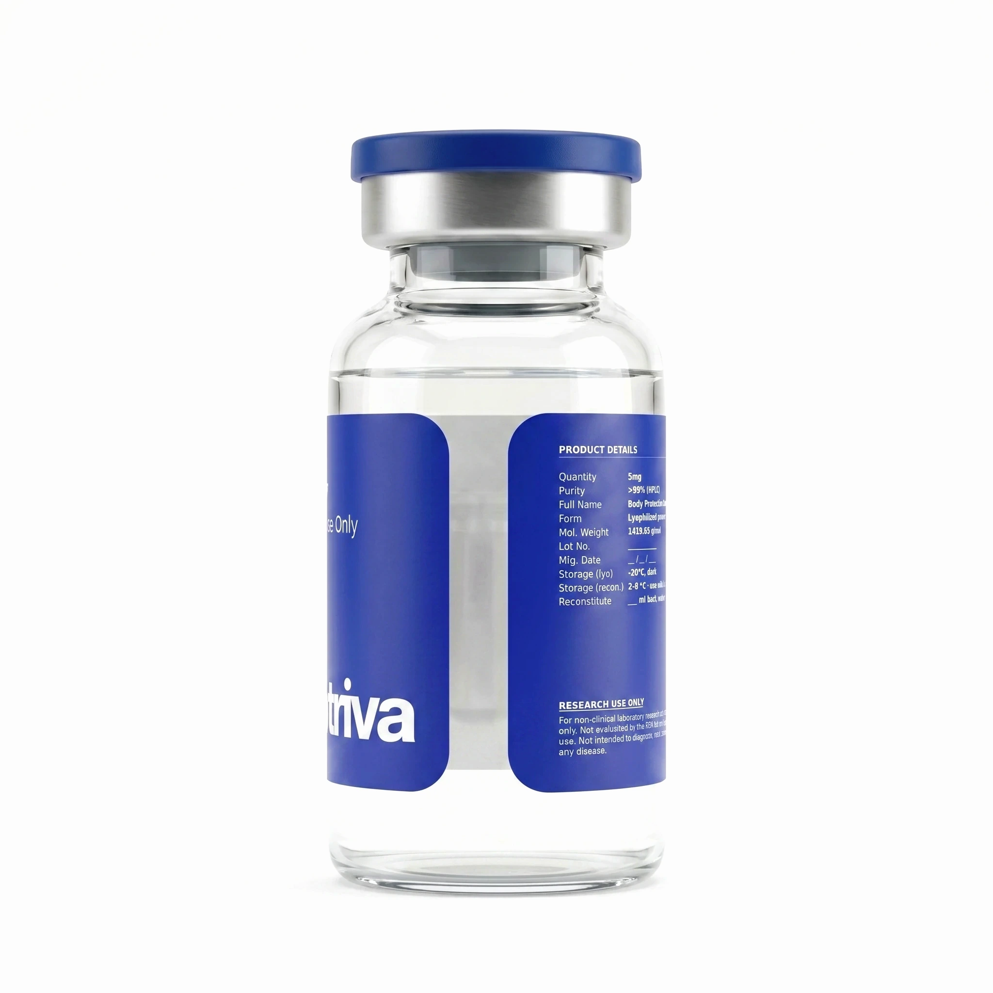
BPC-157
The reference lyophilized vial used to develop the reconstitution protocol in this guide. Lab-verified identity and purity.
What are the common mistakes?
Most reconstitution failures fall into 4 categories. Three of them are technique problems, not equipment problems.
Foaming. The single most common avoidable error. It happens when you blast the diluent straight at the cake, or shake the vial to dissolve it instead of swirling. Foam isn’t cosmetic. Peptides denature at the air-liquid interface, and persistent foam means you’ve probably damaged a real fraction of the molecule. The fix is mechanical. Slow wall-injection, gentle swirl, zero agitation.
Contamination from sloppy stopper technique. Two failure modes show up. You skip the alcohol swab, or you puncture the same spot on the stopper over and over. Both raise the chance of introducing bacteria. The fix is discipline. Swab every time. Vary the puncture point slightly so you don’t weaken one spot.
Volume errors. Misreading the syringe scale. The worst offender is mixing up 100-unit insulin syringes (where 1 mL equals 100 units) with 1 mL tuberculin syringes. Concentration errors there compound through every downstream withdrawal. The fix: pick one syringe type per protocol and check the volume mark before every transfer.
Storage errors. Leaving a reconstituted vial on the counter, or freezing and thawing it repeatedly. Both shorten the use window. Most reconstituted peptides survive a single freeze-thaw if you absolutely need to, but repeated freezing is degrading. Fix: refrigerate immediately after labeling. Minimize temperature swings.
The single biggest predictor of whether a lyophilized peptide vial holds its specification through the in-use window is the technique used in the first three minutes of reconstitution. Wall-injection, gentle swirling, sterile stopper handling. Get those right and the rest is mostly storage discipline.
— American Society of Health-System Pharmacists, Handbook on Injectable Drugs, current edition
What does a properly reconstituted vial look like?
This is the visual checklist worth memorizing. The solution is:
- Clear and colorless (some peptides go slightly straw-yellow at high concentrations, but most are water-clear)
- Free of visible particles
- Not cloudy
- Free of persistent foam after sitting for a minute
- Free of any undissolved cake at the bottom
The stopper should look intact, with no obvious damage from your needle. The label should record the reconstitution date and final concentration in handwriting that’ll survive several days in the fridge.
Where this falls short: when to discard. Toss any reconstituted vial that shows cloudiness, visible particles, unusual color or smell, persistent foam after 1 minute, undissolved cake after 5 minutes of swirling, stopper damage, or any sign of contamination. A tossed vial costs less than a downstream experiment poisoned by a bad reference solution.
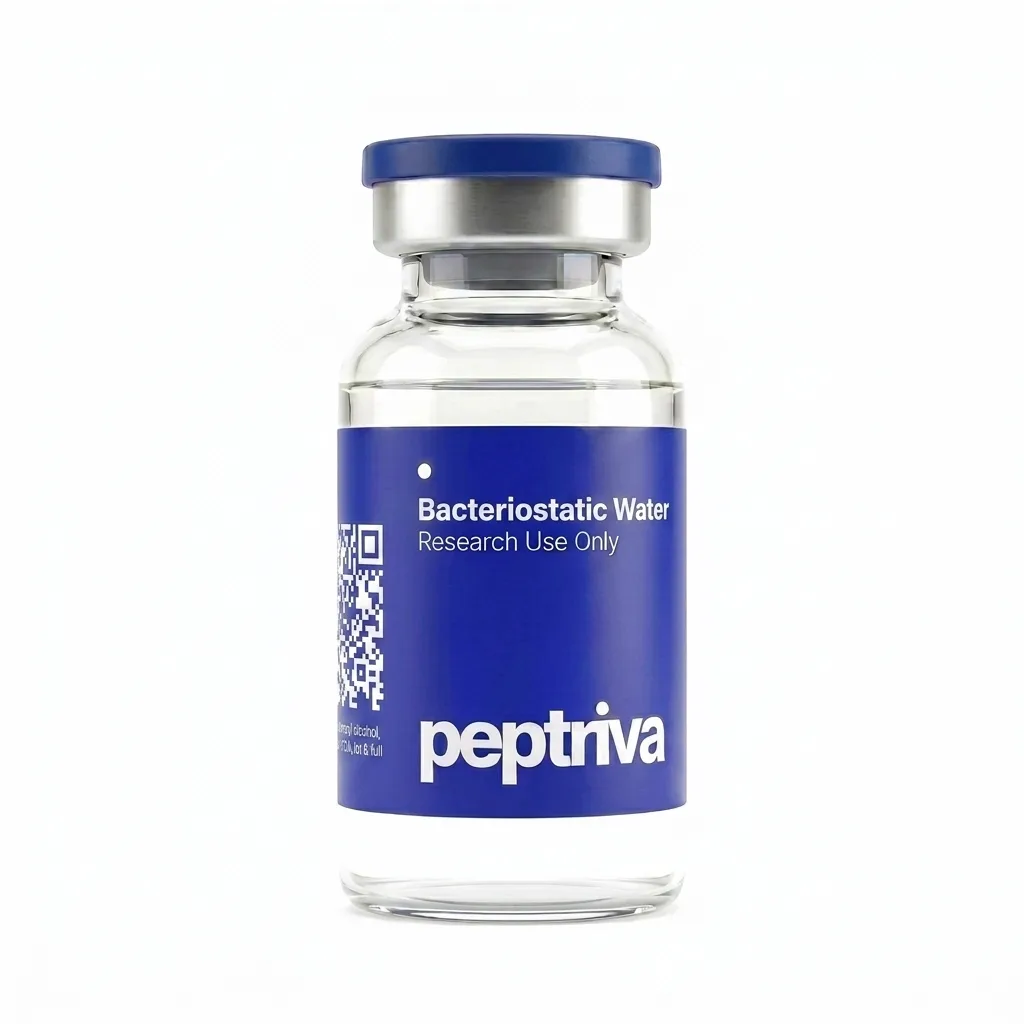
Bacteriostatic Water
Bacteriostatic water · 10 mL sterile vial, 0.9% benzyl alcohol. The diluent used across the reconstitution procedures in this guide. Sealed sterile presentation.
What about storage between uses?
Once you’ve reconstituted and inspected the vial, it lives in the fridge at 2 to 8°C. Most household fridges run in this range. Keep the vial away from the freezer compartment (where temperatures drop below 0°C) and away from the door (where the temperature cycles every time you open it). A small dedicated bin reduces handling errors.
Before each subsequent withdrawal, inspect the vial again for clarity. Let it warm on the bench for 5 to 10 minutes before drawing. Cold solution flowing through a fine needle shears the peptide more than necessary. A short warm-up reduces that stress.
The 28-day window is the standard pharmaceutical convention for a BAC water-reconstituted multi-dose vial. Some peptides have shorter or longer documented stability windows. When the manufacturer provides specific data, follow that data. The general rule: 28 days is the default upper bound, and any visible breakdown (cloudiness, color change, particles) means discard, no matter how far you are into the window.
Key procedural variables that require compound-specific data
Several reconstitution parameters are compound-specific and cannot be generalized. Researchers should confirm these against the compound’s documentation or supplier COA before beginning a protocol:
- Reconstitution volume and target concentration. Volume is compound-specific. The required diluent volume is determined by the peptide mass in the vial and the target working concentration for the assay. There is no universal answer.
- Diluent compatibility. Most peptides are compatible with bacteriostatic water. Some compounds have documented benzyl alcohol incompatibilities and require an alternative diluent (e.g., sterile water for injection or acetic acid).
- Documented in-use stability window. The 28-day convention is a general pharmaceutical baseline for BAC-water multi-dose vials. Individual peptides may have shorter or longer documented stability windows; compound-specific stability data overrides the default where available.
- Clarity acceptance criteria. Clear solution, no foam, intact stopper. The visual inspection is the real-time quality check before each withdrawal.
- Sterile technique requirements. Clean bench, alcohol swabs at every septum puncture, single-use needles, refrigerated storage between uses. All four are required to maintain the in-use window.
What to know now
- Eight-step procedure: temperature equilibration, septum disinfection, diluent draw, wall-injection, gentle swirl, dissolution wait, clarity inspection, label and refrigerate.
- Wall-injection is the key step: diluent runs down the inner glass, never directly onto the cake. This single technique prevents most foaming and denaturation problems.
- Swirl, never shake: peptides denature at air–liquid interfaces; shaking introduces foam and mechanical stress that damage the molecule.
- Bacteriostatic water is the default diluent: 0.9% benzyl alcohol gives a 28-day in-use window at 2–8°C for multi-dose vials.
- Inspect for clarity: the reconstituted solution should be clear, colorless, particulate-free. Anything else is a discard signal.
- Label the date and concentration: the 28-day window starts on the reconstitution date, not the diluent open date.
- Refrigerate, don’t freeze: 2–8°C between uses; minimize freeze–thaw exposures; allow brief warm-up before each withdrawal.
- When in doubt, discard: the cost of a discarded vial is always lower than the cost of using a compromised reference solution.
What we’re watching
Two developments worth tracking. First, the ongoing shift in some specialty pharmaceutical preparations toward single-dose prefilled syringes that eliminate the reconstitution step entirely — useful for clinical settings but largely outside the research-grade context, where lyophilized multi-dose vials remain the standard format. Second, manufacturer-specific stability data for newer peptides — the 28-day in-use convention is a general pharmaceutical baseline, but individual peptides may have shorter or longer documented stability windows that should govern actual practice. Where peptide-specific stability documentation exists, it overrides the default.
References
- United States Pharmacopeia. Bacteriostatic Water for Injection, USP. Monograph in current USP-NF. https://doi.org/10.4135/9781412963855.n1200
- American Society of Health-System Pharmacists (ASHP). Handbook on Injectable Drugs. Bethesda, MD: ASHP, current edition. https://doi.org/10.3109/9780824706081.014
- Trissel, L. A. Handbook on Injectable Drugs. Bethesda, MD: ASHP, current edition. https://doi.org/10.1093/ajhp/46.10.2176
- Wang, W. (1999). Instability, stabilization, and formulation of liquid protein pharmaceuticals. International Journal of Pharmaceutics, 185(2), 129–188. https://doi.org/10.1016/s0378-5173(99)00152-0